Hand and foot numbness can occur for various reasons. It may be due to issues with lumbar or cervical discs, or it could be a result of peripheral neuropathy. In this session, we will explore peripheral neuropathy.
As winter brings chilly winds, many people report experiencing numbness and tingling in their hands and feet. Most often, they attribute these symptoms to poor circulation caused by the cold weather. However, the underlying causes of hand and foot numbness can include circulatory disorders, lower back nerve compression, peripheral neuropathy, spinal disorders, strokes, and even psychological issues.
Peripheral Neuropathy: Tingling Symptoms on Both Sides.
One of the most representative and common causes of hand and foot numbness is peripheral neuropathy. The central nervous system includes the brain and spinal cord, while the thin nerves that branch out from the spinal cord are called peripheral nerves. These peripheral nerves pass through small openings between the vertebrae, known as "nerve foramina," and spread widely throughout the body, including the arms, legs, and torso. They can be categorized into three types based on their function: motor, sensory, and autonomic nerves, with different symptoms arising from damage to each type. Peripheral neuropathy is a condition caused by damage to the sensory nerves of the peripheral nervous system, which spreads throughout the body like electrical wires. If the motor nerves are damaged, weakness and muscle wasting may occur. If the autonomic nerves are affected, it can lead to abnormal sweating, disturbances in bowel and bladder functions, and dizziness. There are various causes of peripheral neuropathy, but damage to the peripheral nerves in the neck and lower back is particularly common. The neck and lower back are areas that experience frequent movement, as the cervical and lumbar vertebrae are continuously moved through connected joints. This movement can lead to chronic compression of the nerve roots passing through the openings between the vertebrae, resulting in damage. This is referred to as "radiculopathy," which is one of the most common causes of hand and foot numbness.
The causes of peripheral neuropathy include:
Diabetes: Diabetic neuropathy, resulting from high blood sugar levels, is the most common cause of nerve damage.
Infections: Viral or bacterial infections, such as shingles, HIV, or Lyme disease, can damage nerves.
Injury: Physical trauma or repetitive stress can lead to nerve damage.
Toxins: Exposure to alcohol, heavy metals, or certain chemicals can negatively impact nerve health.
Autoimmune Diseases: Conditions like rheumatoid arthritis and lupus can affect nerve function.
Nutritional Deficiencies: A deficiency in essential nutrients, particularly vitamin B12, can lead to problems with nerve function.
In the case of polyneuropathy, where multiple peripheral nerves are damaged simultaneously, tingling symptoms typically first appear in the soles of the feet or fingertips. These symptoms gradually progress symmetrically to the entire limbs, often leading to significant pain that can hinder daily activities. This can result in discomfort while walking, running, using chopsticks, or writing.
The symptoms of peripheral neuropathy include:
A burning pain often starting in the hands or feet.
A tingling sensation in specific areas, resulting from nerve damage.
Numbness, particularly commonly felt in the fingertips or toes.
Increased numbness when adopting certain positions.
A loss of sensation, leading to decreased or absent feeling in affected areas.
Muscle weakness, making it difficult to lift objects.
Easy fatigue during daily activities and a lack of energy.
A loss of balance, resulting in instability when walking or standing.
An increased risk of falling or injury.
Changes in temperature sensation, leading to dulled or heightened responses to normal temperatures.
Skin color changes, which may appear red or pale.
Altered sweating, resulting in either reduced sweating or excessive sweating.
Difficulty performing fine motor tasks, such as writing or buttoning clothes.
Symptoms may appear symmetrically on both sides or may affect only one side.
Symptoms often worsen at night, potentially disrupting sleep.
Variability in symptoms throughout the day, with increased fatigue.
Discomfort in daily life, which can lead to mental stress.
As symptoms progress, muscle atrophy or abnormal reactions may occur.
A decreased sensitivity to various stimuli, which may result in unrecognized minor injuries or wounds.
"Neglecting peripheral neuropathy can lead to the progression of numbness throughout the body and may even result in paralysis," he stated. "If you experience new-onset numbness in the soles of your feet or the tips of your fingers that gradually worsens, along with difficulties in walking or using chopsticks, it's important to confirm the presence of peripheral neuropathy through tests such as electromyography (EMG), nerve conduction studies, and evoked potentials."
If Your Fingers Are Numb, Suspect Carpal Tunnel Syndrome
In peripheral neuropathy, mononeuropathy occurs when peripheral nerves are compressed by surrounding structures such as bones, joints, ligaments, and muscles. This type of neuropathy typically presents with numbness limited to one arm or one leg, with carpal tunnel syndrome being the most representative condition.
Carpal tunnel syndrome results from compression of the nerve in the wrist due to the wrist ligaments and joint. It primarily manifests as numbness in the thumb, index, and middle fingers, often worsening after extensive use, but symptoms may improve when shaking the hand.
"If symptoms such as pain and numbness occur when the wrist is fully flexed, or if light tapping over the middle area of the wrist elicits numbness, carpal tunnel syndrome should be suspected," he stated.
Cervical Disc Herniation: Causes Shoulder Pain and Arm Numbness
If you experience numbness in your hands accompanied by a tingling sensation in your fingers, shoulder pain, headaches, or stiffness in the back of your neck, there is a high possibility that it could be due to a cervical disc herniation (cervical intervertebral disc herniation). In this condition, the nucleus inside the disc between the cervical vertebrae protrudes and compresses the nerve roots or spinal cord, leading to symptoms such as stiffness and aching in the neck, as well as numbness and pain in the shoulders, arms, and hands. Additionally, if you experience numbness on the sides or back of your legs, it may indicate a lumbar disc herniation, and if you have numbness in the ankles, calves, thighs, or buttocks accompanied by lower back pain, spinal canal stenosis should be considered.
"Even if numbness is felt in the hands or feet, the actual cause is often related to spinal disorders in the cervical or lumbar regions," he noted. "Especially in cases where a patient has undergone spinal surgery or has chronic neck and back pain, the cause of numbness in the hands and feet is likely to be a spinal condition."
Q1. If peripheral neuropathy is suspected, which department should I visit at the hospital?
For detailed diagnostic tests and appropriate treatment, it is recommended to consult a neurologist. However, if extensive testing is unnecessary or if you only need symptom management, it is also acceptable to see a physician with relevant experience or consult in the departments of orthopedics, neurosurgery, pain medicine, or rehabilitation medicine.
Disc calcification refers to the condition where calcium deposits accumulate in the annulus fibrosus or the central part of the intervertebral disc, causing the disc to become hard and bone-like.
Causes
Disc calcification can be caused by degenerative changes related to aging, trauma, or genetic factors. Disc protrusion occurs when the nucleus pulposus, the central part of the disc, pushes through the annulus fibrosus. As the disc protrudes, inflammation can combine with calcium deposits, leading to disc calcification.
Since disc calcification forms over a long period, it often appears in individuals who have had disc problems for an extended time. Additionally, inflammation caused by disc removal surgery can contribute to calcification, which is why post-surgery care to prevent inflammation is crucial.
Symptoms
Disc calcification can cause lower back pain, and if the calcification presses on a nerve, it may result in radiating pain in the legs. The symptoms may vary depending on the location of the calcification and which nerve is compressed..
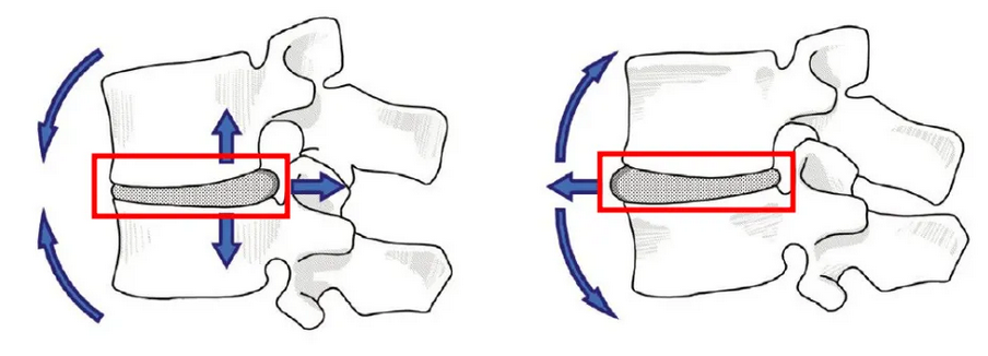
At the area where bones meet cartilage, there is a structure known as the endplate (A), which has high elasticity and absorbs the impact on the disc.
The nucleus pulposus (B) and annulus fibrosus (C) do not contain blood vessels, making them unable to heal once damaged.
(A = Endplate, B = Nucleus Pulposus, C = Annulus Fibrosus)
Disc Protrusion
When a disc is subjected to pressure, the part that typically gets damaged is the outer layer known as the annulus fibrosus.
Pain begins when the annulus fibrosus is compromised.
Continuously bending forward causes the annulus to thin and the nucleus pulposus to dehydrate.
Eventually, this leads to disc protrusion, as shown in the illustration below.
When the nucleus pulposus tears through the annulus fibrosus, it creates an injury, leading to the death of nucleus pulposus cells and the production of inflammatory substances.
This process results in pain, known as discogenic pain, which tends to be localized around the lower back.
Disc Rupture
When the limits of disc protrusion are exceeded, the dehydrated nucleus pulposus tears through the annulus fibrosus, resulting in a disc rupture.
A disc rupture can cause excruciating pain that is often unimaginable.
Severe Radicular Pain
When the injury to the annulus fibrosus worsens, it becomes thinner, and the nucleus pulposus also dehydrates, causing the nucleus to tear through the annulus and escape.
This condition is referred to as a disc rupture. When a disc ruptures, it can compress the spinal cord or nerve roots, leading to leg numbness or weakness.
Additionally, the inflammatory substances released from the escaping nucleus can irritate the nerve roots. When these nerve roots are compressed or stretched, severe pain radiates down the leg, known as radicular pain.
Radicular pain typically starts in the lower back and travels through the buttocks, thigh, calf, and down to the foot. The intensity of this pain is often beyond imagination.
When the nucleus pulposus escapes through the annulus fibrosus, the inflammatory substances released from the damaged nucleus flow towards the nerve roots.
Natural Healing
When the nucleus pulposus ruptures, it releases inflammatory substances.
However, while these inflammatory substances cause pain, they also play a role in healing the torn annulus fibrosus. This is similar to the natural healing process of skin wounds..
Disc degeneration refers to a chronic pain condition caused by damage to the internal structure of the intervertebral disc. This condition occurs when the disc, located between the vertebrae, experiences internal damage or degeneration without herniating outward. It is considered one of the main causes of lower back pain. Disc degeneration is related to the aging process or degenerative changes in the spine, particularly when the annulus fibrosus (A) is damaged, leading to increased pressure within the disc.
Structure and Function of the Disc
The intervertebral disc is a structure located between the vertebrae that absorbs shock and allows for flexibility in spinal movement. The disc consists of two main parts:
Nucleus pulposus: Located at the center of the disc, this jelly-like substance serves to absorb shock.
Annulus fibrosus: A tough fibrous tissue that surrounds the nucleus pulposus, protecting the disc from external impacts.
Disc degeneration primarily results from damage to the annulus fibrosus. Unlike herniated discs, which involve problems occurring externally, disc degeneration is characterized by issues arising internally. (A = Annulus fibrosus, B = Nucleus pulposus)
Causes of Disc Degeneration
Disc degeneration primarily occurs due to the following reasons:
Aging: As people age, the discs naturally degenerate, losing their elasticity and becoming more susceptible to damage.
Repetitive Stress: Lifting heavy objects repeatedly or applying continuous pressure on the back can lead to disc damage.
Poor Posture: Sitting for long periods or working in awkward positions can increase the load on the discs, resulting in damage.
Trauma: Injuries from accidents, falls, or other traumatic events can damage the discs.
Symptoms
The main symptom of disc degeneration is chronic lower back pain, which typically has the following characteristics:
Localized Back Pain: Pain is primarily concentrated in the lower back, with radiating pain to the legs or buttocks being less common.
Increased Pain When Sitting: Sitting or changing positions can exacerbate the pain.
Pain When Bending Forward: Bending the back forward may intensify the pain.
Persistent Pain: The pain generally lasts for several weeks and can sometimes continue for months.
Diagnosis
To diagnose disc degeneration, the following methods are employed:
Medical History and Physical Examination: The doctor will listen to the patient's symptoms and check spinal movement and the location of the pain.
Imaging Tests: MRI (Magnetic Resonance Imaging) scans can reveal the internal structure of the disc and identify any damage. A CT discography, which involves injecting dye into the disc, may also be used to identify the source of pain.
Treatment
Treatment for disc degeneration is divided into conservative and surgical approaches.
Conservative Treatment:
Medications: Nonsteroidal anti-inflammatory drugs (NSAIDs) or muscle relaxants may be used to alleviate pain.
Physical Therapy: Includes posture correction, exercise therapy, and strengthening exercises to reduce the burden on the disc by strengthening surrounding muscles.
Injection Therapy: Treatments such as nerve blocks or steroid injections may be used to reduce pain.
Surgical Treatment:
Endoscopic Discectomy: In cases of severe disc damage, surgery may involve removing or stabilizing the damaged disc using an endoscope.
Artificial Disc Replacement: This involves replacing the damaged disc with an artificial one, but this surgery is only suitable under specific conditions.
Prevention
Posture Correction: Maintaining proper posture while sitting and standing is essential.
Exercise: Regular stretching and strengthening exercises for the back can help reduce stress on the discs.
Weight Management: Maintaining a healthy weight is beneficial for spinal health.
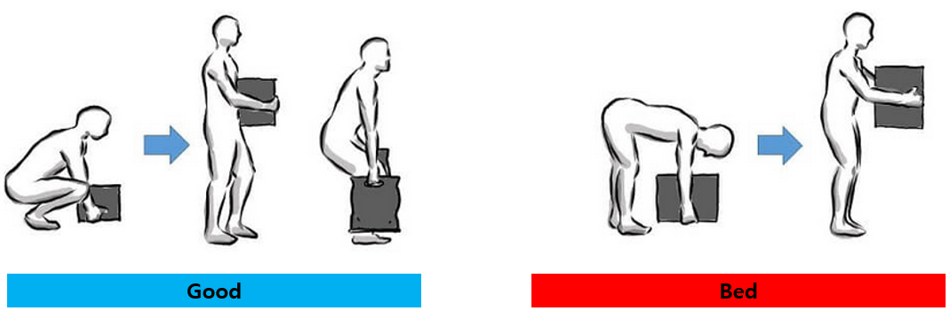
Proper Lifting Techniques: Instead of bending at the waist, it's better to bend at the knees when lifting heavy objects to reduce strain on the discs.
Disc degeneration can be managed effectively through appropriate treatment and care, helping to alleviate pain and improve quality of life..
1. Incision in the Back Skin 2. Removal of the Lamina or Facet Joint using a Drill (Irreversible)
3. Removal of the Ligamentum Flavum (Irreversible) 4. Repositioning the Spinal Cord with Surgical Instruments (Avoiding Direct Contact)
The endoscopic surgical instruments will enter in the direction of the red arrow. If there are difficulties in removing the nucleus due to disc rupture, additional bone removal may be necessary. In some cases, a significant amount of bone from the facet joints may be removed. Therefore, it is advisable to discuss with your doctor how much bone will be removed before the surgery.
Cauda Equina Syndrome (CES) is a neurological emergency that occurs at the lower spine, primarily caused by conditions such as spinal stenosis, herniated discs, trauma, or tumors. It involves the compression of the "cauda equina," a bundle of nerves located below the lumbar vertebrae. Symptoms can include lower back pain, bilateral leg pain and sensory changes, muscle weakness, altered sensation in the perineal area, and dysfunction in bowel and bladder control. If not treated promptly, CES can lead to severe complications.
1. Causes
Herniated Disc: A spinal disc displaces and compresses nearby nerves.
Spinal Stenosis: Narrowing of the spinal canal leads to nerve compression.
Trauma: Damage from accidents or falls can result in CES.
Tumors: Tumors within the spine can exert pressure on the nerves, causing CES..
2. Symptoms
Lower Limb Paralysis: Reduced mobility or paralysis in the lower body.
Sensory Changes: Loss of sensation or abnormal sensations (tingling, numbness) in the buttocks, thighs, and legs.
Bowel and Bladder Issues: Dysfunction may lead to incontinence or difficulty in controlling urination and defecation.
Sexual Dysfunction: Problems with sexual function, which may lead to decreased sexual pleasure.
3. Diagnosis
Physical Examination: Neurological assessments and symptom verification are conducted.
Imaging Tests: MRI or CT scans are used to identify the cause of nerve compression.
Electrophysiological Testing: Tests assess nerve function and determine the extent of damage.
4. Treatment
CES is a neurological emergency, so early intervention is critical.
Surgery: Surgical intervention may be necessary to relieve nerve compression, commonly through discectomy or laminectomy.
Medications: Steroids or pain relievers can be used to reduce inflammation.
Physical Therapy: Rehabilitation may be required post-surgery.
5. Prognosis
If treated promptly, there is a high likelihood of symptom improvement. However, delaying treatment can lead to permanent nerve damage or disability. Therefore, it is crucial to seek medical attention immediately if symptoms of CES occur.
Cauda Equina Syndrome can significantly affect the nervous system, making accurate diagnosis and treatment essential. Quick response to symptoms is important to prevent complications.
6. Example of Cauda Equina Syndrome
Below, in Figure 1, the left side shows an image of a herniated disc.
Cauda Equina Syndrome only occurs when the nerve bundle is compressed; thus, in cases like that shown in Figure 1, CES cannot develop.
Below, in Figure 2, there is an image showing a herniated disc in the central region.
In this case, the herniated disc is compressing the nerve bundle, which can lead to the development of Cauda Equina Syndrome.
7. Conclusion
As illustrated in the example above, Cauda Equina Syndrome is likely to develop only when the nerve bundle in the central region is compressed by a herniated disc. If an MRI shows that a disc, as in Figure 2, is compressing the nerve bundle and bowel or bladder dysfunction is present, surgery is recommended as an emergency.
Additionally, if surgery is delayed while the nerve remains compressed for an extended period, significant time may be required for nerve recovery.
Therefore, it is advisable to head directly to the emergency room if there are issues with bowel or bladder function.
8. Q / A
Q: If an MRI shows a disc protrusion to the left without compressing the central nerve bundle but bowel and bladder dysfunction is present, what should be considered?
A: The presence of bowel and bladder dysfunction indicates that there may be nerve compression occurring elsewhere, not necessarily at the site of the disc protrusion. For example, a tumor in the thoracic spine could compress the nerve bundle, or there could be compression from another area of the lumbar spine. It may be beneficial to undergo a full-body MRI scan to investigate further.
Maintaining good habits and proper posture is essential for preventing herniated discs. Since we spend a significant amount of time sleeping and sitting, it's important to maintain good posture in these activities. There are many things to consider for spinal health and preventing back pain, such as driving, household chores, and lifting objects. Let’s explore what to do and what to avoid in daily life to prevent herniated discs.
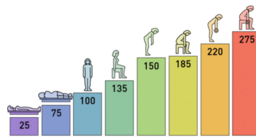
Disc pressure according to various postures
Lifting Objects
When lifting objects from the floor, adopt a squatting position by bending your knees while keeping your back and upper body as upright as possible. Then, as you stand up, straighten your knees while using the strength of your legs rather than your back. It is very important to avoid bending at the waist while straightening your legs to lift the object. This technique helps protect your spine and prevent strain on your back.
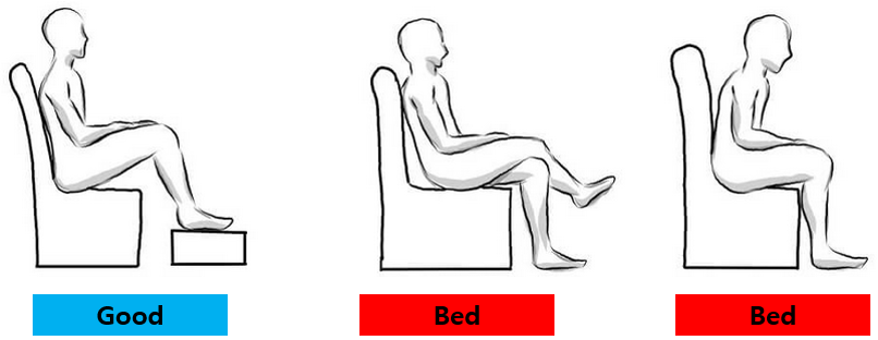
Sitting Posture
It is not advisable to sit at the edge of the chair with your hips pushed back or to cross your legs while sitting. You should also avoid slouching or leaning forward. Always try to sit in a chair that has back support, and if possible, use a chair with armrests for added comfort and support.
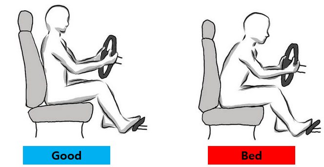
Driving Posture
Tilt the backrest of the driver's seat about 10 degrees backward and sit with your lower back and buttocks firmly against the seat. It's ideal to keep your upper body upright at a 90-degree angle; however, instead of tensing your muscles to maintain this position, lean back against the seat for a more relaxed posture, which puts less strain on your back. Avoid leaning forward while driving or sitting at the edge of the seat.
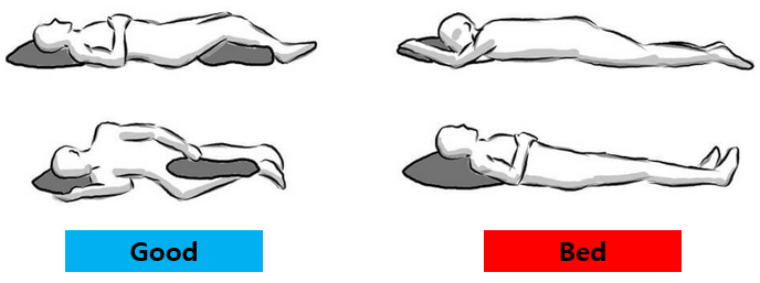
Sleeping Posture
Sleeping on your back or on your side is generally acceptable, but it's best to avoid sleeping on your stomach whenever possible.
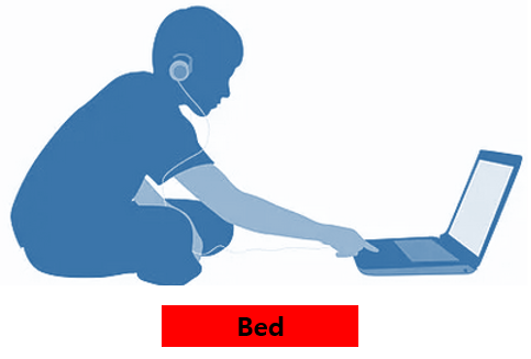
Sitting on the Floor
When sitting on the floor, it’s common for the back to lean forward, which can put strain on the spine. Therefore, it’s best to avoid sitting directly on the floor if possible.
Brushing Posture
When brushing your teeth, avoid bending at the waist and use a cup for rinsing. Spit into the cup, then rinse your mouth with water from the cup after cleaning it.
Head Washing Posture at the Sink
Do not bend your head down to wash your hair at the sink. Instead, wash your hair while standing in the shower.
Face Washing Posture at the Sink
Avoid leaning over the sink when washing your face. It’s better to wash your face while showering.
Household Chores
For daily tasks like cleaning and washing dishes, maintain good posture. Avoid squatting to sweep or mop the floor. Use a vacuum cleaner or a mop with a handle. When using these tools, adjust the handle length to suit your height, keeping your back straight and your body upright. Standing while bending at the waist is not good for your back.
Footwear
High-heeled shoes and flat shoes with a thickness of less than 1 cm are not beneficial for back health. Opt for shoes that have a flat sole, with a heel height of about 2-3 cm, and provide some shock absorption while walking. Height-increasing insoles are also not good for your back.
Chair and Bed Habits
It’s better to sleep in a bed and sit in a chair rather than sleeping on the floor or sitting on the floor. When sleeping in a bed, avoid mattresses that are too hard or too soft.
Exercises Beneficial for the Back
Recommended exercises for preventing herniated discs include walking, cycling, swimming, and yoga. Riding a bike can strengthen the muscles in your hips and lower body, which support your back. However, avoid bikes that require excessive bending at the waist, and be cautious of riding on uneven surfaces. The use of an indoor bike with back support is preferable.
Swimming is highly beneficial for preventing herniated discs if you are healthy and fit. However, be cautious with strokes like the butterfly or breaststroke. Stretching and core-focused exercises like yoga and Pilates are also excellent for back health.
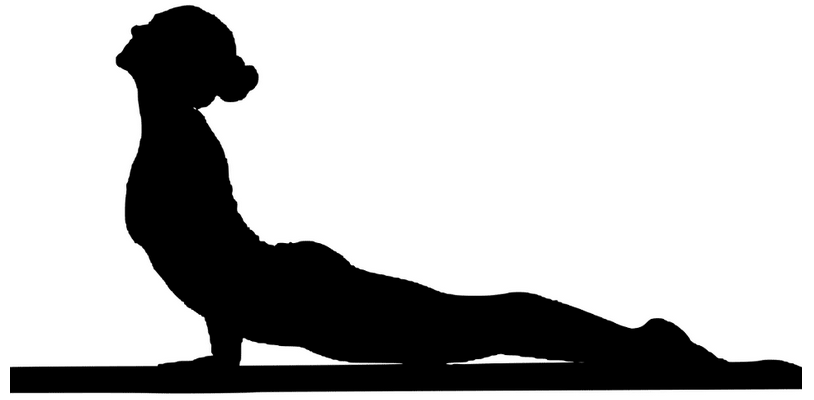
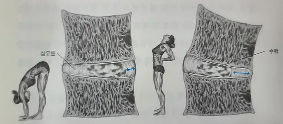
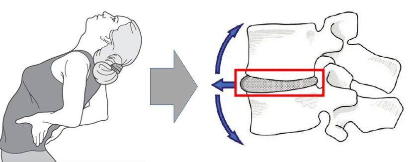
Importance of Lumbar Lordosis Posture
Lumbar lordosis refers to the curvature of the lumbar spine (lower back). When lifting something from the floor or maintaining a hunched position, pressure can be placed on the intervertebral discs, leading to back pain. In these situations, maintaining lumbar lordosis is effective in alleviating discomfort.
When maintaining lumbar lordosis posture, the torn inner fibers of the intervertebral discs come into contact with each other, promoting recovery. Furthermore, when the natural curve of the lower back is maintained, the discs can withstand more than ten times the load compared to a flat back, helping to prevent disc damage. Therefore, If you maintain a forward-leaning posture, the muscles in your lower back will weaken and naturally return to the original posture.